
As we continue training into our 40s, 50s, and 60s, we occasionally notice a “gap” between numbers and bodily sensations. Body weight remains largely unchanged. Muscle mass measured by body composition monitors does not show a drastic decline. Yet, we might feel that “we cannot produce force as explosively as we used to,” “explosive output in sprints or on hills has dropped,” or “fatigue lingers differently than it used to after the same training.”
In explanations of conventional “muscle aging,” such changes tend to be attributed to “muscle mass decreasing due to aging.” Of course, the age-associated reduction in muscle mass is an important phenomenon. However, changes in muscle mass alone cannot explain everything.
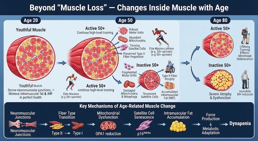
Age-related changes in skeletal muscle involve multiple factors, including not only muscle fiber atrophy and loss, but also neuromuscular changes, fiber-type-specific changes, intramuscular fat, mitochondrial function and quality control, and the regenerative environment including muscle stem cells. Wilkinson et al. reviewed evidence showing that age-related muscle loss involves both muscle fiber atrophy and the loss of individual muscle fibers, with multiple mechanisms—including changes in motor units and reduced responsiveness to physical activity and nutrition—contributing to these processes.
In other words, when we feel our “muscles have declined,” the aspect changing is not necessarily just the “amount of muscle.”
Here, another important issue arises. What we call “age-related muscle decline” may include not only changes due to age per se, but also the effects of long-term physical activity levels or inactivity. Individuals who have exercised for many years and those who have aged with little physical activity do not necessarily have the same muscle condition, even at the same age.
Muscle is a tissue that cannot be captured solely by quantitative measures such as muscle mass.
- 1: The Explanation of “Muscle Loss” Leaves Something Missing
- 2: Muscle Is Not a “Lump of Flesh,” But an Assembly of Multiple Systems
- 3: Muscle Performance Is Not Determined by Size Alone
- 4: The State of Mitochondria Also Changes Within Muscle
- 5: “Repair and Adaptation” Are Supported by Muscle Stem Cells and Immune Cells
- 6: “Age-Related Changes” and “Inactivity-Related Changes” Are Not the Same
- 7: To What Extent Can Long-Term Exercise Maintain the Muscle Environment?
- 8: Muscle Has a “History”
- 9: Considering “What We Want to Maintain” in Training from Age 50
- 10: Shifting Focus from “Muscle Mass” to “Actual Function”
- Conclusion:Looking at “Contents” and “Time,” Not Just Muscle “Quantity”
- References
1: The Explanation of “Muscle Loss” Leaves Something Missing
First, let us consider the “muscle mass” figures we commonly encounter. Home body composition monitors and devices using bioelectrical impedance analysis (BIA) are convenient tools for estimating body composition based on the electrical characteristics of the body. However, the “muscle mass” displayed there is not a value obtained by directly extracting and weighing muscle tissue.
Therefore, even if the numbers for muscle mass are relatively maintained, it does not automatically mean that muscle function is maintained just as it was in youth. Herein lies the difference between “quantity” and “function.”
In age-related skeletal muscle changes, not only muscle fiber atrophy occurs, but also the loss of muscle fiber itself. Furthermore, changes in the nervous system that control muscle movement, such as the reduction and reorganization of motor units, are known to occur.
In other words, “a decrease in muscle mass” and “a decline in muscle function” are not completely identical phenomena.
A single numerical value like muscle mass cannot evaluate muscular strength, power, neural recruitment, metabolic function, and tissue composition all at once. That is why considering age-related muscle changes requires not only a quantitative perspective of “how many kilograms exist,” but also a perspective of “how that muscle is functioning.”
The studies discussed in this article do not all address the same aspect of muscle aging. By viewing different studies side by side—encompassing muscle fibers, the neuromuscular system, intramuscular fat, mitochondria, and muscle stem cells—we examine muscle changes that cannot be captured by the single number of muscle mass alone.
2: Muscle Is Not a “Lump of Flesh,” But an Assembly of Multiple Systems
Skeletal muscle is not a simple chunk of protein. For muscles to generate force, utilize energy, adapt to training stimuli, and repair damaged tissue, numerous components must work in coordination.
Inside muscle, there are elements such as the following:
- Muscle fibers: Generate force through contraction
- Motor neurons and motor units: Innervate muscle fibers and control contraction
- Mitochondria: Produce ATP (energy)
- Blood vessels and capillaries: Transport oxygen and nutrients
- Connective tissue: Supports the structure of muscle tissue
- Muscle satellite cells (muscle stem cells): Involved in the regeneration of muscle tissue
- Immune cells: Involved in processes of damage and regeneration
Regarding muscle regeneration, it is known that the surrounding microenvironment, including immune cells such as macrophages as well as muscle satellite cells, plays a crucial role. The review by Wang and Zhou describes how macrophages and muscle satellite cells interact in a coordinated manner during muscle regeneration following injury.
Importantly, these components do not all change at the same rate.
- Even if muscle mass is relatively maintained, the neuromuscular system may have undergone changes.
- Even if muscle fiber size has not changed significantly, the distribution of intramuscular fat may have altered.
- Not just the amount of mitochondria, but the state of proteins involved in their morphology and function may have shifted.
Considered this way, a single term like “muscle aging” is clearly insufficient to fully express the changes actually taking place.
3: Muscle Performance Is Not Determined by Size Alone
Crucial to considering muscle function are the muscle fibers themselves and the nervous system that recruits them. Particularly in age-related changes, the atrophy of Type II fibers (so-called fast-twitch muscle fibers) can be prominent.
Verdijk et al. (2007) compared 8 elderly men around 76 years of age with 8 young men around 20 years of age, examining muscle fibers in the vastus lateralis. As a result, the elderly subjects showed a smaller proportion and average cross-sectional area of Type II fibers compared to the young subjects, along with a lower satellite cell count per Type II fiber. On the other hand, similar reductions in satellite cell counts were not confirmed in Type I fibers.
This study demonstrated an association between Type II fiber atrophy and reduced satellite cell content in the elderly. However, it was a comparative study of 8 versus 8 subjects and did not simply prove that “the reduction of satellite cells caused Type II fiber atrophy.”
Additionally, aging induces changes in the motor units that innervate muscle fibers. The review by Wilkinson et al. summarizes evidence for human muscle fiber loss and motor unit reduction, suggesting that structural changes in aged muscle are related to the reorganization of denervated muscle fibers by surviving motor neurons.
What emerges from this is that muscle function is not simply determined by “muscle size.” Muscle fiber types and sizes, the motor units innervating the fibers, and their reorganization combine to determine actual force-generation capacity.
Given this background, the concept of “dynapenia” becomes important. Dynapenia refers to the age-related loss of muscle strength and power that is not fully explained by a reduction in muscle mass.
Therefore, muscle strength decline should not be explained by a single cause such as “because muscles became smaller,” but rather considered comprehensively, including muscle fibers, the neuromuscular system, and muscle quality.
Intramuscular Fat
When considering the internal characteristics of muscle, another factor that cannot be overlooked is intramuscular fat (IMF).
Yoshiko et al. (2017) used MRI to three-dimensionally investigate thigh intramuscular fat in 15 young adults around age 21 and 15 elderly adults around age 70. In the elderly, the thigh muscle cross-sectional area was smaller than in the young, and in some regions, the proportion of intramuscular fat relative to muscle mass was higher. Meanwhile, the cross-sectional area of intramuscular fat itself showed little difference between young and old adults in certain regions.
In other words, the increase in the intramuscular fat proportion observed in parts of this study was partly due to the relative proportion becoming higher because muscle mass decreased, rather than a massive absolute increase in fat itself. Therefore, it is inappropriate to oversimplify this as “aging causes massive accumulation of fat inside muscles.”
However, the composition and distribution of muscle and fat can provide additional information about muscle quality and function beyond what muscle mass alone can tell us. This is yet another change difficult to perceive through the single figure of “muscle mass.”
4: The State of Mitochondria Also Changes Within Muscle
For muscles to be active, large amounts of ATP are required. Mitochondria play a central role in supplying this energy. However, when considering mitochondria, looking merely at “whether there are many or few” is insufficient.
Mitochondria repeatedly undergo fusion and fission within cells, altering their morphology and function. Such “mitochondrial dynamics” represent an important mechanism for maintaining mitochondrial function. Furthermore, “mitophagy”—the selective degradation of damaged mitochondria—is one of the key mechanisms of mitochondrial quality control.
However, studies must not be confused here.
What the OPA1 Study by Tezze et al. Showed
Tezze et al. (2017) focused on “OPA1,” a protein related to mitochondrial morphology and function. In humans, OPA1 levels were lower in sedentary older individuals, whereas this age-related reduction was less pronounced in older individuals who remained physically active.
Furthermore, in mice, muscle-specific deletion of Opa1 revealed pathways leading to altered mitochondrial morphology and function, alongside ER stress, FoxO-mediated catabolic signaling, and muscle atrophy.
In other words, the key takeaway from this study is that “mitochondria are not mere ATP factories, but the maintenance of their morphology and function itself relates to muscle homeostasis.”
On the other hand, treating this paper as a “study on mitophagy” is inappropriate. OPA1 is a protein related to mitochondrial fusion and morphological maintenance, and the core of Tezze et al.’s study lies in OPA1 reduction and muscle, metabolic, and systemic changes. Therefore, we do not explain that “Tezze et al. proved that reduced mitophagy causes muscle atrophy.” Mitochondrial dynamics and mitophagy are related quality control systems, but they are not the same thing.
Mitochondrial DNA Abnormalities and Muscle Fibers
Wanagat et al. (2001) examined aged rat muscle in detail, reporting that regions containing mitochondrial DNA deletion mutations co-localized with electron transport system abnormalities, muscle fiber atrophy, fiber splitting, and oxidative damage.
However, this study did not target humans; the subjects were Fischer 344 × Brown Norway F1 hybrid rats. Consequently, it cannot be concluded that mitochondrial DNA deletions directly cause muscle fiber damage in humans.
What can be said from this study is that in aged rat muscle fibers, mitochondrial DNA deletion mutations, electron transport system abnormalities, and muscle fiber atrophy existed in the same regions. As an animal study, it serves as important material for considering age-related muscle changes at the cellular level, but animal study results cannot be directly substituted for human phenomena.
5: “Repair and Adaptation” Are Supported by Muscle Stem Cells and Immune Cells
Muscles possess mechanisms to respond to damage and regeneration. One of the key players is the muscle satellite cell (muscle stem cell).
Muscle satellite cells normally reside in a quiescent state. When stimuli such as muscle injury occur, they activate, proliferate, and differentiate, supporting the repair and regeneration of muscle fibers. However, muscle repair is not completed by satellite cells alone.
The review by Wang and Zhou (2022) organizes how, during the regeneration process following muscle injury, immune cells such as macrophages interact with muscle satellite cells to participate in inflammation, clearance of damaged tissue, regeneration, and tissue environment remodeling. In other words, muscle “repair” is not a simple mechanism where a single cell type fixes broken muscle, but a complex process in which multiple cells and signals overlap temporally.
Age-Related Changes Also Occur in Muscle Satellite Cells
Functional changes in muscle satellite cells due to aging have also been studied.
Research by Sousa-Victor et al. (2014) showed that $p16^{\text{INK4a}}$ expression increases in muscle satellite cells of aged mice, making cells that are normally in a reversible quiescent state more prone to shifting toward a senescent state. The study also showed that suppressing $p16^{\text{INK4a}}$ restored regenerative capacity in satellite cells from aged mice.
However, caution is needed here as well. This study is primarily a foundational study targeting muscle stem cells in aged mice. Therefore, it cannot be directly concluded that “50-year-old humans have delayed recovery after training because their muscle satellite cells are senescent.”
Age-related changes in muscle stem cells represent important biological background for understanding muscle regenerative capacity, but actual post-training fatigue and recovery times in humans involve various factors, including sleep, nutrition, training load, muscle damage, neuromuscular fatigue, body temperature, and hydration status.
Thus, it is not a simplistic story of “aged muscle becoming unable to repair.” More accurately, it should be considered that “age-related changes also occur in the cells and environments supporting muscle repair and regeneration.”
6: “Age-Related Changes” and “Inactivity-Related Changes” Are Not the Same
Reviewing the research covered thus far reveals that age-related muscle changes are not a single phenomenon.
- Changes in Type II fibers
- Reduction and reorganization of motor units
- Distribution of intramuscular fat
- Mitochondrial morphology and function
- Age-related changes in muscle stem cells
- Regenerative environment including immune cells
These are phenomena investigated by different studies. Furthermore, another crucial factor is “differences in physical activity levels.”
Within the group labeled “elderly,” some individuals have barely moved their bodies for many years, while others have continued exercising for decades. Ignoring this distinction and lumping “elderly muscle” together confounds the effects of age per se with the effects of physical activity and inactivity.
Venturelli et al. (2014) compared young individuals, active elderly individuals, and inactive elderly individuals, examining skeletal muscle telomere length and oxidative stress-related indicators. As a result, differences were observed in cell senescence-related indicators between elderly individuals maintaining physical activity and inactive elderly individuals.
The study therefore suggests that physical inactivity may be an important contributor to cellular aging in skeletal muscle, but it does not establish that aging itself has no effect.
Accordingly, writing off everything with “it’s just age, so it can’t be helped” is inappropriate. On the other hand, claiming “if you exercise, you don’t age” is also incorrect.
A more realistic view is that “changes due to aging itself and changes governed by physical activity levels overlap to shape the current state of muscle.”
7: To What Extent Can Long-Term Exercise Maintain the Muscle Environment?
Regarding how long-term physical activity affects muscle, research has been conducted in animals as well as humans.
White et al. (2016) used C57BL/6J mice to have them perform voluntary resistance wheel exercise for 34 weeks starting from mid-life, subsequently examining their muscles. As a result, age-related muscle loss was attenuated in certain hindlimb muscles examined in the exercise group, and hypertrophy was observed in the soleus muscle. Furthermore, changes were seen in mitochondrial density, indicators of oxidative capacity, and autophagy-related indicators such as the LC3-II/I ratio.
What can be learned from this study is that “resistance exercise continued from mid-life contributed to the maintenance of muscle mass and muscle metabolism-related indicators in aged mice.”
However, this is ultimately mouse research. Moreover, because autophagy-related indicators like the LC3-II/I ratio changed, this cannot be directly expressed as “mitophagy was activated.” Autophagy and mitophagy are related concepts, but they are not identical. Therefore, specific conclusions cannot be drawn from this study to state to humans that “performing this exercise activates mitochondrial mitophagy and prevents aging.”
Even so, it serves as material for considering that long-term exercise stimuli relate not merely to muscle size, but also to muscle metabolism and intracellular environment.
8: Muscle Has a “History”
Looking at the preceding research piece by piece reveals that understanding muscle requires another important time axis: what is referred to here as “muscle history.”
However, let us clarify one thing first. The term “muscle history” is not used here as an established technical term from a specific paper. It is an organizing concept used in this article to collectively express the time axis of “what kinds of physical demands and training stimuli the muscle has experienced over time” in order to understand the current state of muscle.
Two people are in their 50s and weigh the same. Their estimated muscle mass by body composition monitors is also nearly identical. Even so, it does not mean their muscles are in the same state. A person who has exercised continuously since their 20s and a person who barely exercised for years and started exercising only after turning 50 have experienced vastly different stimuli, even if their current muscle mass is similar.
Of course, this does not mean that “having exercised for years keeps all muscle in the same state as a youth.” Age-related changes remain. However, maintaining physical activity over the long term can preserve parts of muscle structure and metabolic function more favorably, as can be considered from the studies introduced so far.
- Tezze et al.’s study: Mitochondrial-related factors such as OPA1 are maintained in active elderly individuals.
- White et al.’s mouse study: Continuous resistance exercise from mid-life maintained muscle mass and mitochondrial/autophagy-related indicators in old age.
- Other studies: Reports indicating age-related changes in muscle satellite cells and Type II fibers also exist.
In short, muscle neither “does not age if you exercise” nor “inevitably declines in the same way as you age.” Current muscle is formed as the result of various cumulative influences over a long time, including aging, physical activity, inactivity, nutrition, disease, and training. This time axis is what this article calls “muscle history.”
9: Considering “What We Want to Maintain” in Training from Age 50
As we have seen, muscle is not a tissue with a single function.
- Maintaining muscle mass
- Generating force
- Producing force rapidly
- Supplying energy over long durations
- Adapting to stimuli
- Repairing damaged tissue
These are not entirely identical mechanisms. That is precisely why training does not need to be considered solely through the single objective of “increasing muscle mass.”
For example, it can generally be organized as follows:
| Function to Maintain/Improve | Principal Exercise Stimulus |
| Muscle mass & foundational strength | Resistance training |
| High force and power output | Short-duration high-output exercise and power training |
| Aerobic capacity & metabolic function | Endurance exercise |
| Overall muscle function | Combining multiple stimuli while allowing adequate recovery |
However, this is not a correspondence table stating that “performing this exercise directly improves this cellular function.”
For instance, performing strength training does not automatically improve the nervous system, muscle fibers, mitochondria, and muscle stem cells all in the same way. Nor does endurance exercise guarantee sufficient maintenance of functions responsible for high output.
What matters is not treating muscle as a single unit, but applying different stimuli to different functions according to objectives. Furthermore, as one ages, viewing not just the training itself, but also the extent of recovery achieved following training, becomes crucial.
There is no need to establish blanket rules such as “high intensity only once a week because I’m in my 50s.” The important thing is to observe how one’s body actually responds to actual training loads.
10: Shifting Focus from “Muscle Mass” to “Actual Function”
Let us return these discussions back to daily training.
The numbers on a body composition monitor certainly serve as one point of reference. However, muscle status cannot be fully judged solely by how many kilograms of muscle mass are present. Therefore, we also focus on daily bodily changes:
- Whether force can be generated rapidly on hills or in sprints, just like before
- Whether necessary output can be maintained during high-intensity exercise
- Whether the body works stably during long-duration exercise
- How conditioning recovers after training compared to before
- Whether performance is maintained or improved when continuing the same training
Such actual functions are key.
Of course, such bodily sensations alone cannot accurately diagnose the internal state of muscle. It is impossible to determine things like “legs feel heavy, so mitochondria are bad” or “muscle soreness lasts long, so muscle stem cell function has declined.” This is because daily performance and fatigue are influenced by many factors, including sleep, nutrition, training load, psychological stress, body temperature, and hydration status.
Even so, when training is followed under reasonably consistent conditions over a long period, observing how the body actually performs can provide information that a single measure of muscle mass cannot capture.
Conclusion:Looking at “Contents” and “Time,” Not Just Muscle “Quantity”
From the phrase “muscle aging,” one might imagine muscle simply being chipped away little by little. However, much more complex changes occur within actual skeletal muscle.
- Muscle fibers atrophy
- Some muscle fibers are lost
- Motor units decrease and reorganize
- Changes appear in Type II fibers
- The proportion and distribution of intramuscular fat alter
- Shifts occur in mitochondrial morphology and function
- The properties of muscle stem cells change
- The regenerative environment including immune cells also changes
And these changes involve not only age, but also the long time axis of how much physical activity and training the body has experienced over the years.
That is why “how many kilograms of muscle mass exist” cannot fully represent a person’s current muscular state. At the same time, there is no need to think “aging is not occurring because muscle mass is maintained.” Even when muscle mass is relatively maintained, age-related changes can occur in muscle fibers, the neuromuscular system, and the metabolic system.
Conversely, just because one has aged, there is no need to resign oneself to all muscle changes as “inevitable aging.” Continuing physical activity may suppress some age-related changes or maintain muscle function. However, those effects vary depending on the research subjects and exercise methods, and do not mean all aging phenomena can be prevented.
When examining muscle, it is important to hold three perspectives:
- “How many kilograms are present right now?”
- “What is its structure, and how is it functioning?”
- “What kind of stimuli has it received up to now?”
“Muscle history,” as termed in this article, is one organizing method for considering that time axis. The muscles of someone who has exercised for many years also change with age, but those muscles exist here now as the result of stimuli received over decades.
Therefore, what matters in training from age 50 is not simply “recovering the muscle of youth.”
Understanding the functions that the current body possesses, maintaining remaining capabilities, providing appropriate stimuli to necessary functions, and ensuring sufficient recovery. By looking beyond muscle mass to the changes taking place inside muscle and to its history, our relationship with an aging body should look a little different.
References
- Verdijk et al. (2007)
Verdijk LB, Koopman R, Schaart G, Meijer K, Savelberg HHCM, van Loon LJC. Satellite cell content is specifically reduced in type II skeletal muscle fibers in the elderly. Am J Physiol Endocrinol Metab. 2007;292(1):E151-E157. PMID: 16926381. PubMed - Wilkinson et al. (2018)
Wilkinson DJ, Piasecki M, Atherton PJ. The age-related loss of skeletal muscle mass and function: Measurement and physiology of muscle fibre atrophy and muscle fibre loss in humans. Ageing Res Rev. 2018;47:123-132. PMID: 30048806. PubMed - Yoshiko et al. (2017)
Yoshiko A, Hioki M, Kanehira N, Shimaoka K, Koike T, Sakakibara H, Oshida Y, Akima H. Three-dimensional comparison of intramuscular fat content between young and old adults. BMC Med Imaging. 2017;17(1):12. PMID: 28183279. PubMed - Tezze et al. (2017)
Tezze C, Romanello V, Desbats MA, et al. Age-Associated Loss of OPA1 in Muscle Impacts Muscle Mass, Metabolic Homeostasis, Systemic Inflammation, and Epithelial Senescence. Cell Metab. 2017;25(6):1374-1389.e6. PMID: 28552492. PubMed - Wanagat et al. (2001)
Wanagat J, Cao Z, Pathare P, Aiken JM. Mitochondrial DNA deletion mutations colocalize with segmental electron transport system abnormalities, muscle fiber atrophy, fiber splitting, and oxidative damage in sarcopenia. FASEB J. 2001;15(2):322-332. PMID: 11156948. PubMed - Sousa-Victor et al. (2014)
Sousa-Victor P, Gutarra S, García-Prat L, et al. Geriatric muscle stem cells switch reversible quiescence into senescence. Nature. 2014;506:316-321. PMID: 24522534. PubMed - Wang & Zhou (2022)
Wang X, Zhou L. The Many Roles of Macrophages in Skeletal Muscle Injury and Repair. Front Cell Dev Biol. 2022;10:952249. PMID: 35898401. PubMed - Venturelli et al. (2014)
Venturelli M, Morgan GR, Donato AJ, et al. Cellular aging of skeletal muscle: telomeric and free radical evidence that physical inactivity is responsible and not age. Clin Sci (Lond). 2014;127(6):415-421. PMID: 24708050. PubMed - White et al. (2016)
White Z, Terrill J, White RB, McMahon C, Sheard P, Grounds MD, Shavlakadze T. Voluntary resistance wheel exercise from mid-life prevents sarcopenia and increases markers of mitochondrial function and autophagy in muscles of old male and female C57BL/6J mice. Skeletal Muscle. 2016;6:45. PMID: 27964759. PubMed
コメント